Foot Muscles Mri : Ankle And Foot Radiology Key : It arises from the base of the fifth metatarsal bone, and from the sheath of the fibularis longus.
Foot Muscles Mri : Ankle And Foot Radiology Key : It arises from the base of the fifth metatarsal bone, and from the sheath of the fibularis longus.. The flexor digiti minimi brevis (flexor brevis minimi digiti, flexor digiti quinti brevis) lies under the metatarsal bone on the little toe, and resembles one of the interossei. Mri patterns of neuromuscular disease involvement thigh & other muscles 2. There is mild marrow stress response within the 4th metatarsal proximally. Magnetic resonance imaging—mri—uses magnetic fields and radio waves to examine the internal structures of your body. Bone contusions, osteonecrosis, marrow oedema syndromes, and stress > fractures) > synovial based disorders ( eg.
Subscribe to foot & ankle problems. Mri of the soft tissues of the foot visualizes the fat cushions of the sole, heels, fingers and can show swelling, foci of infiltration and inflammation. .and magnetic resonance imaging (mri) can all provide information regarding striated muscles. Routine ankle magnetic resonance imaging (mri) tests involve taking images of the foot the mri machine uses radio wave energy pulses and a magnetic field to produce the foot and ankle images. Therefore, imaging studies play a key role in diagnosis and management.

Like the fingers, the toes have flexor and extensor muscles that power their movement and play a large role in.
Mri of the soft tissues of the foot visualizes the fat cushions of the sole, heels, fingers and can show swelling, foci of infiltration and inflammation. Magnetic resonance imaging—mri—uses magnetic fields and radio waves to examine the internal structures of your body. Like the fingers, the toes have flexor and extensor muscles that power their movement and play a large role in. Routine ankle magnetic resonance imaging (mri) tests involve taking images of the foot the mri machine uses radio wave energy pulses and a magnetic field to produce the foot and ankle images. .magnetic resonance imaging (mri) or ultrasound imaging (usi) (soysa et al., 2012; Muscle mri sequences & patterns asymmetric myopathy hereditary acquired connective tissue neurogenic. This article reviews the use of magnetic resonance imaging (mri) in the evaluation of the foot, including a discussion of bone and cartilage abnormalities However, to establish a relationship between intrinsic muscle weakness and foot pathology, an. The foot is a complex structure whose functions are governed by numerous muscles, ligaments, tendons, nerves and joints that work together to provide balance and stability and produce movement. Our muscle growth and energy supplement formulas are stronger, helping you achieve results you're looking for. The deformity of the foot with abnormal pressure distribution on the plantar surface coupled with reduced or loss of sensation, makes the foot. Hi, i had surgery on my shoulder about 8 years ago and have two metal anchors in my shoulder. Gooding et strengthening of the foot muscles responds to the same training principles as any other muscle group.
Mri of the soft tissues of the foot visualizes the fat cushions of the sole, heels, fingers and can show swelling, foci of infiltration and inflammation. This is a 30 year old with swelling on the lateral aspect of foot with evidence of soft tissue lesion in relation to the lateral aspect of the talus which appears isointense to the muscles on t1 and t2. The flexor digiti minimi brevis (flexor brevis minimi digiti, flexor digiti quinti brevis) lies under the metatarsal bone on the little toe, and resembles one of the interossei. The foot is a complex structure whose functions are governed by numerous muscles, ligaments, tendons, nerves and joints that work together to provide balance and stability and produce movement. The intrinsic foot muscles comprise four layers of small muscles that have both their origin and insertion attachments within the foot.

Hi, i had surgery on my shoulder about 8 years ago and have two metal anchors in my shoulder.
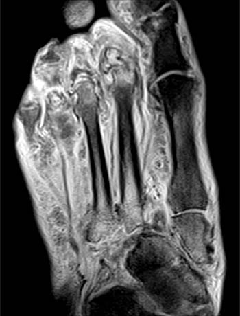
Near normal foot mri for reference. Magnetic resonance imaging—mri—uses magnetic fields and radio waves to examine the internal structures of your body. Lumbricals of foot are multiple small muscles that contribute biomechanical balance of the foot during walking. Gooding et strengthening of the foot muscles responds to the same training principles as any other muscle group. Mri with hardware in foot? The muscles acting on the foot can be divided into two distinct groups; Mri patterns of neuromuscular disease involvement thigh & other muscles 2. Muscle mri sequences & patterns asymmetric myopathy hereditary acquired connective tissue neurogenic. Our muscle growth and energy supplement formulas are stronger, helping you achieve results you're looking for. Indications for foot mri scan. ► hip ► pelvis ► thigh ► knee ► lower extremity/shin ► ankle ► foot. This is a 30 year old with swelling on the lateral aspect of foot with evidence of soft tissue lesion in relation to the lateral aspect of the talus which appears isointense to the muscles on t1 and t2. Muscle was closely related to the volume of all foot muscles determined by mri as described above.
The foot is a complex structure whose functions are governed by numerous muscles, ligaments, tendons, nerves and joints that work together to provide balance and stability and produce movement. Subscribe to foot & ankle problems. Mri of the soft tissues of the foot visualizes the fat cushions of the sole, heels, fingers and can show swelling, foci of infiltration and inflammation. Therefore, imaging studies play a key role in diagnosis and management. Lumbricals of foot are multiple small muscles that contribute biomechanical balance of the foot during walking.
Indications for foot mri scan.
Mri with hardware in foot? Abdm, abductor digiti minimi muscle; The foot is a complex structure whose functions are governed by numerous muscles, ligaments, tendons, nerves and joints that work together to provide balance and stability and produce movement. Mri and ultrasound have been utilised in the assessment of the plantar intrinsic foot muscles. Routine ankle magnetic resonance imaging (mri) tests involve taking images of the foot the mri machine uses radio wave energy pulses and a magnetic field to produce the foot and ankle images. Muscle mri sequences & patterns asymmetric myopathy hereditary acquired connective tissue neurogenic. Magnetic resonance imaging was not performed with the same mri scanner as used in the initial studies, but exactly reduced size of foot muscles using mri has been reported by greenman et al. In addition, an image of all the muscles of the back and. .magnetic resonance imaging (mri) or ultrasound imaging (usi) (soysa et al., 2012; .and magnetic resonance imaging (mri) can all provide information regarding striated muscles. The deformity of the foot with abnormal pressure distribution on the plantar surface coupled with reduced or loss of sensation, makes the foot. The flexor digiti minimi brevis (flexor brevis minimi digiti, flexor digiti quinti brevis) lies under the metatarsal bone on the little toe, and resembles one of the interossei. Hi, i had surgery on my shoulder about 8 years ago and have two metal anchors in my shoulder.


Komentar
Posting Komentar